What Are The Two Types Of Leukemia – How many types of leukemia are there? What makes each of them unique and treated differently?
To answer these questions and more, we turned to leukemia specialist Naveen Pemmaraju. Here’s what he shared.
Contents
- 1 What Are The Two Types Of Leukemia
- 2 Nelarabine, Intensive L Asparaginase, And Protracted Intrathecal Therapy For Newly Diagnosed T Cell Acute Lymphoblastic Leukaemia In Children And Young Adults (all T11): A Nationwide, Multicenter, Phase 2 Trial Including Randomisation In The Very High Risk
- 3 Blood Cancer: What Are The Various Types And Treatments Available?
What Are The Two Types Of Leukemia

A little bit. Leukemia is not just a disease. This is actually a different group of blood-based cancers that originate in the bone marrow.
What Is Leukemia: Symptoms, Causes & Treatment
Examples of younger patients might be an athlete getting lab work before knee surgery, or someone getting blood tests to get new life insurance. Young patients with chronic leukemia often have no symptoms, so they are usually diagnosed incidentally.
Yes It is further divided by the type of white blood cells involved: either myeloid or lymphocytes. The four main types of leukemia are:
Philadelphia-positive leukemia is characterized by translocations between chromosomes 9 and 22. It occurs when part of chromosome 9 breaks and joins with chromosome 22, causing abnormal cells to form.
Philadelphia-positive was one of the first types of leukemia to prove that it could be treated with oral chemotherapy, and MD Anderson’s leukemia group was the first to combine this approach with immunotherapy, leading to very high cure rates. Interestingly, the Philadelphia chromosome is present in 100% of CML cases, but is also present to a lesser extent in AML and ALL.
Nelarabine, Intensive L Asparaginase, And Protracted Intrathecal Therapy For Newly Diagnosed T Cell Acute Lymphoblastic Leukaemia In Children And Young Adults (all T11): A Nationwide, Multicenter, Phase 2 Trial Including Randomisation In The Very High Risk
Some types of leukemia are rare or rare, but each of them has developed its own research area for different reasons found. Thanks to our unique approach to treatment, many patients can live with some of these subtypes for decades. Our overall goal for all of these diseases is to maximize patient response while improving their quality of life. Leukemia is the most common cancer in children. Morphological analysis of bone marrow smear is an important first step in diagnosis. Recent publications have shown that AI can differentiate between blood cells, but is far from clinical use. A total of 1732 bone marrow images were used to train a convolutional neural network (CNN). New deep learning techniques were combined and a complete leukemia diagnosis system was developed using raw images without pre-processing. The system creatively mimics the workflow of a hematologist, identifying and removing uncountable and weakened cells, then isolating and counting the remaining cells for diagnosis. The performance of CNN in WBC classification achieved 82.93% accuracy, 86.07% accuracy and 82.02% F1 score. And the accuracy of diagnosis of acute lymphoid leukemia reached 89%, sensitivity 86% and specificity 95%. The system also performs well in detecting bone marrow metastases in lymphoma and neuroblastoma, achieving an average accuracy of 82.93%. This is the first study involving a variety of different cell types to diagnose leukemia with high performance in real clinical settings.
Leukemia, which results from the arrest of nucleated cell growth and differentiation, which can interfere with normal blood cell production, can develop at any age from newborns to the elderly and is most devastating, mainly in children, accounting for up to 30% of patients. all childhood cancers (1). In addition, these immature cells can spread through the blood and invade other organs, causing multiple organ dysfunction and ultimately death. Due to the rapid proliferation and spread of leukemia cells, early and accurate diagnosis is urgently needed.
Despite the widespread use of immunological, cytogenetic and molecular tests, morphological analysis of bone marrow nodules is still an important initial step in the diagnosis of leukemia (2, 3) because it is an economical and convenient method. Pathologists analyze the characteristics of blood cells, such as shape, size, and granularity, to determine cell types using a light microscope, and then perform tests as directed. However, classical morphological diagnosis is tedious and labor-intensive work that requires time and highly trained specialists, and the results of diagnosis can be subjective. On the other hand, computer-aided diagnosis (CAD) system helps to save time and overcome the shortcomings of manual work, including fatigue, substance and so on.

The basis of the morphological diagnosis of leukemia is the differential count of white blood cells (WBC). Computational analysis based on deep learning has shown potential promise as a strategy for identifying distinct populations of WBCs. Choi et al. (4) and Qin et al. (5) demonstrated the ability of deep learning to classify white blood cells at different stages of development, which makes a deep diagnosis based on leukemia, but these studies had limitations related to small cell types and accuracy. Below was the classification usually . created using pre-processed images, not raw medical images. WBC differential count in bone marrow analysis is an important application of deep learning, but needs improvement.
Blood Cancer: What Are The Various Types And Treatments Available?
In this study, bone marrow images of children with leukemia were collected from Shanghai Children’s Medical Center and white blood cells were described. We used the results to create a leukemia cell database called the AI Cell Platform. Cell images from the data were used for training and testing to develop a leukemia diagnostic system that used depth to distinguish up to 19 types of white blood cells at different stages of development in real clinical settings, instead of using pre-processed images or data. public on the Internet. . Different types of leukocytes were able to fulfill the criteria for the diagnosis of childhood cancer. Our system mimics the bone marrow analysis process of hematologists. In addition, we further evaluated the feasibility of an AI system for the diagnosis of acute lymphoblastic leukemia (ALL) in clinical practice.
To enable the development of machine learning algorithms for diagnosis, we at Shanghai Children’s Medical Center (SCMC) created a database, an AI cell platform, containing 1,732 images obtained from the bone marrow of 89 children with leukemia between 2009 and 2019. The included bone marrow smears were stained according to the Wright-Giemsa protocol. Images of the prepared slides were obtained with a light microscope at 1000x magnification. For each smear, 15 nonoverlapping outlets were averaged and randomly selected. The SCMC Institutional Review Board waived the need for informed consent. All images will be de-identified before they are made available. The images used in this study were produced with a camera (MooGee; 505 C GS).
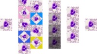
Corresponding diagnoses of each bone marrow specimen were confirmed by flow cytometry. The exact types of white blood cells in each image were described by 2 hematologists who have worked together for over 10 years and checked each other’s description to ensure accuracy. The database consisted of 19 classes of WBC and neuroblastoma cells of different growth stages (Figure 1). In fact, there are about 40 types of white blood cells in the bone marrow, and there are very few specific types of white blood cells. We did not get enough images of these rare WBCs to train the model. Therefore, we combine these cells into a group (class 20) during the training of the classification model. The number of cells in each class and their distribution are shown in Figure 2 and Table 1. The distribution between classes is unbalanced. Since the natural distribution of white blood cells is unbalanced, this problem was inevitable.
The differential WBC counting system consisted of two parts: a diagnostic mode and a classification mode. Raw bone marrow images were first processed in the diagnostic section, where all white blood cells were found in red blood cells, platelets, contaminated sediments, and so on. The identified cells were then used as input to the sorting unit. The classification part consisted of two stages. In the first step, we distinguished uncountable cells, including damaged cells, damaged cells, and so on, which are not used to diagnose leukemia. In the second step, countable WBCs were differentiated in a multistep manner (Figure 3).
Biphenotypic Acute Leukemia: A Report Of Two Cases
Figure 3. The general structure of the system consists of two parts: a recognition module and a classification module. In terms of detection, we train a detection model using the RetinaNet method to detect all white blood cells from bone marrow images. The classification part consists of two stages. In a first step, we will develop a countable cell sorting model to distinguish white blood cell destruction that hematologists would not count. After the second step, the identified countable white blood cells are transferred to the WBC sorting system.
We used RetinaNet (6) VGG (7) and Feature Pyramid Network (8) for cell detection. RetinaNet is a single-phase detector that outperforms all existing two-phase detectors. Supplementary Figure 1 shows the RetinaNet model. The network consists of a backbone network and two sub-networks. The main mesh is used for feature computation and the subnets are used to connect the box backend and classification. FPN is considered a backbone network. It includes upstream downstream (VGG) and reverse link. This structure can create a multidimensional feature pyramid from the input image. Each FPN level has two subnets associated with it: classification
What are the two types of arthritis, what are the types of leukemia, two types of leukemia, what are the 4 types of leukemia, what are the different types of leukemia, what different types of leukemia are there, what are the 2 types of leukemia, what are the two types of lawyers, two kinds of leukemia, what types of leukemia are curable, what types of leukemia are there, what are the different types of leukemia in adults


